The petrographic microscope is used to study
microscopic preparations of soils. This is a microscope that uses
plane polarised light. The samples are placed between two polaroid
plates, whose vibration directions are perpendicular. The minerals
display numerous optical properties in the polarising microscope,
which are very useful for studying them and which are not shown
in a normal or biological microscope.
The lower polaroid is always inserted in the direction of the
light rays and it is called a polarizer. The upper polaroid is
used to analyse the effects that are produced when the polarised
light crosses the minerals and that is why it is called an analyser.
Unlike the polarizer, the analyser is not fixed in the rays direction,
as it can be placed or removed whenever required. It is used for
some specific properties (in this case the term used is crossed
polaroids) but not for others (only polarizer).
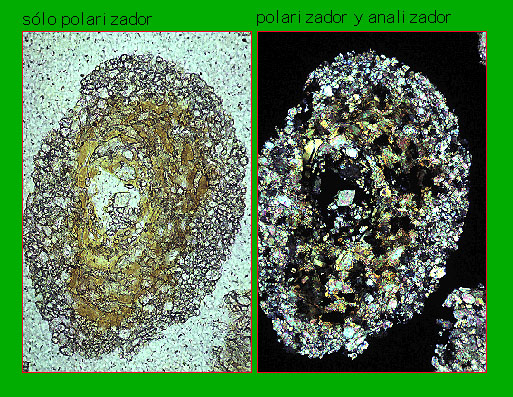
With the polarizer, the view is similar to the one obtained using
a biological microscope with normal light, in other words not
plane polarised light. But with the analyser the situation is
totally different. The minerals appear with artificial colours
(also called interference colours) that are the result of interference
from waves that have suffered from the double refraction in the
mineral. Interference colours help to identify the minerals, to
differentiate different crystals in the same mineral, depending
on the position they have fallen into (it distinguishes polycrystalline
grains from monocrystalline grains) and to recognise the degree
of alteration they have.
The voids in the soil (and the gaps between the sand grains) that we can see in the microscope are filled with an isotropic substance that is used to include the soil samples and give them the coherence required to be able to cut and trim them in order to make microscopic preparations. This substance (it is a liquid polyester resin that hardens on polymerisation), as it is isotropic, does not interfere with the light rays and the voids are seen as black with the crossed polaroids (light rays cannot pass between two polarised plates, whose vibration directions are perpendicular). That is why the photographs taken with only the polarizer appear with a white background, while those with a black background are those taken with crossed Nicols.
In order to differentiate the voids from the opaque minerals (black), the polaroids are sometimes placed with their different vibration directions forming angles that are slightly different from 90º (oblique polaroids). In this situation, the voids look grey.
Sometimes microphotographs are taken with the microscope light switched off and using an external light source on the microscope slide. These conditions, known as oblique incidental light or inclined reflected light, are very different from the usual conditions of the microscope in which one works with light transmitted through the minerals.
The scale has been omitted in the photographs of this monograph,
since we are only dealing with coarse sand grains and we know
that their real size is always 2-0.2 mm.
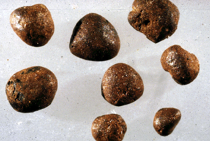
Macrophotographs
Sometimes photos taken with a binocular
loupe are shown. Their reproduction scale is around 1:1 (natural
size), varying between 10:1 and 1:10 (smaller enlargement than
that of the microphotographs). Below there are two macrophotographs
of a granitic rock.