![]() Spanish version
Spanish version

Soil Micromorphology |
|||||||
|
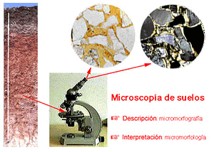 2. Micromorphography Soil description under the microscope |
 3. Micromorphology Soil interpretation under the microscope |
|||||
Carlos Dorronsoro
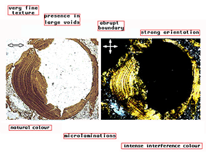
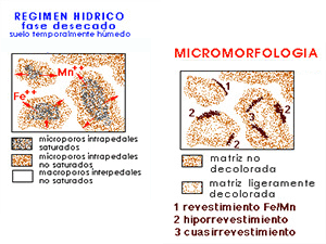
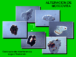
Traditionally, one or two microphotographs are attached to soil micromorphology studies in order to better understand some specific features, but they can hardly provide a global idea of the thin section. We open this section where large microscopic images are shown, so that you can browse them with different zooms (similar to Google Maps/Earth) just as you are actually looking through the petrographic microscope.
only polarizing |
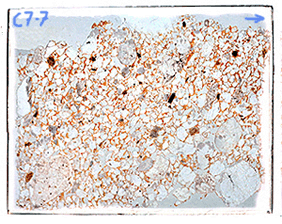
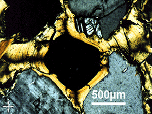
GigaMICROimages are images taken from a very important area of the thin section. They are mosaics composed of 100 to 300 microphotographs taken with the polarizing microscope with different lenses: x2,5, x4, x5, x8 y x10. The size of each microscopic field (mosaic cell) ranges from 2,6x5,5mm to 0,6x0,9mm, depending on the different zooms. The total dimension of the covered area in the microscopic preparations varies between 21x16mm y 53x31mm. The size of resulting digital image differs from 1 to 3 GB but the ones we are browsing have been reduced to 50%. Browsing these huge images would require long loading times, but this problem is overcame using a particular viewer (krpano) that splits up large images into pieces and loads them only when needed. |
|
|
two images attached |
|
|
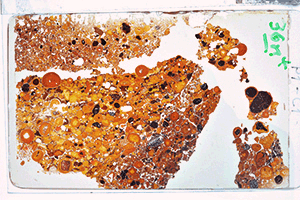
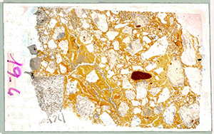
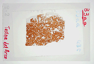
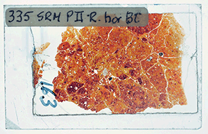
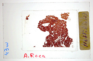
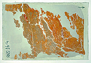
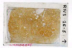
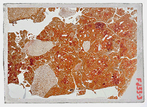
GigaMACROimages are macro images directly taken from the complete thin sections using a flat scanner equipped with a module for films. It is astonishing the information obtained from these images, especially when the thin sections of the soil is located between two acetate polaroid layers that act as polarizer and analizer. The images shown here were taken with a low/medium quality commercial scanner. We think that, if we would had used a scanner for high quality films, the resolution and zoom view of the images would have highly increased. This system can also be useful for images that do not fit in the microscope field even when using the smallest zoom lens. |
two crossed polarizers |
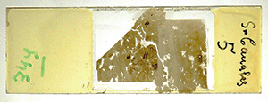
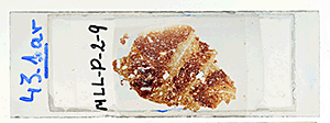
Click on a preparation for the gigaimage.
|
|
|
|
 Bt Horizon Illuviation in very clayey hor. Medium/high magnifications |
|
|
|
|
|
|